
OUR TOOLS
Imaging
This includes imaging techniques using:
- Confocal microscopy
- Intravital microscopy
- Clearing whole tissue

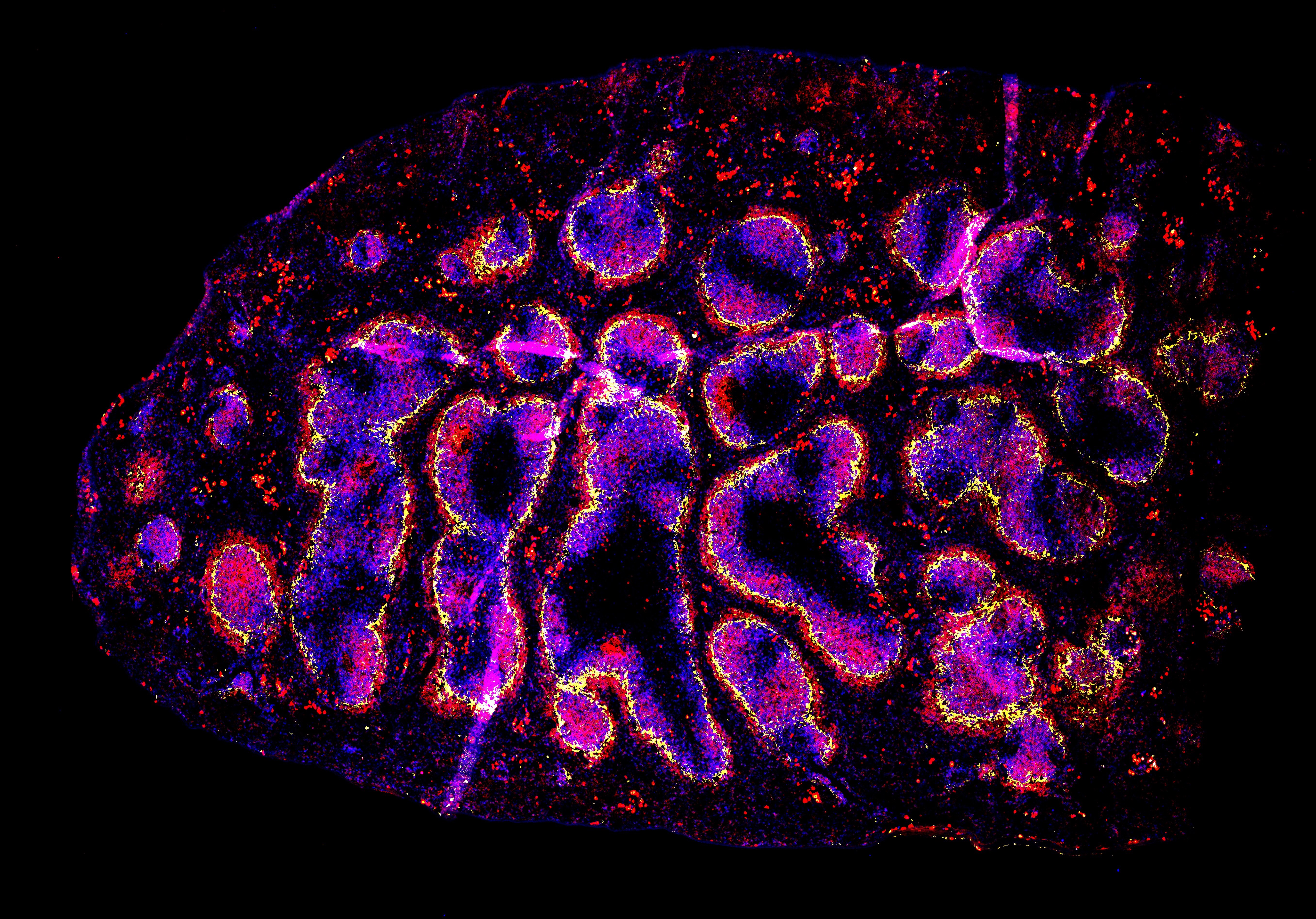
Images of the Spleen
Image credit: Connie Shen
")
Section of cleared tissue (cervical lymph node)
Image credit: Jeremy Postat
In Vitro Migration Assays
This includes:
- Under agar
- Microchannels
- Collagen matrices

Image example of microchannels
> Migration and constriction of Lifeact-GFP+ T cells can be observed in the above image. The red represents Hoechst-labelled nuclei. (Image credit: Jeremy Postat)
Systems Approaches
This includes:
- Single cell sequencing
- RNA sequencing
- TCR sequencing